Immunopath
 | Flexible protocol based image analysis for Whole Slide Images- Immunopath automises laborious and repetetive pathology tasks
- Full image analysis and sampling options for data reduction
- Flexible options for Whole Slide Image support, import from standard imaging devices and common file formats
- Protocol based image analysis for repeatable and reliable results
- Runs on standard desktop PCs or Notebooks
- Also available; Pathtool - a partner application providing tools for grading microscopy images in pathology
|
Human review of slides prepared using Immunohistochemistry techniques can be time consuming and laborious. Counting cells or estimating the percentage stain on a slide is not something the average pathologist enjoys. Our IHC quantification tool offers a protocol based system that can rapidly complete counting and field fractions type quantification from images obtained from standard microscopy cameras, as well as whole slide imaging systems.
Immunohistochemistry (IHC) is a test used by pathologists to detect specific molecules on cells.
When a tissue sample is passed to a lab to be examined for disease, there are several details that cannot be determined easily. Several diseases or disease sub-types may look alike or appear to have similar size cells under a microscope but have different behaviors and necessary treatments. One way to differentiate them is to detect specific molecules on these cells that act as markers.
Immunohistochemistry is a technique that uses antibodies that can seek out, identify and attach themselves to these markers on cells. The antibodies themselves can be seen under the microscope and quantfied.
IHC is used for numerous applications in medicine, especially in cancer diagnosis and drug discovery.
The system
ImmunoPath is designed to provide quantitative information on images obtained from Immunohistochemistry (IHC) images captured using slide scanners and from single images captured with standard microscopy cameras.
The system has been designed so that it is protocol based, initially these can be setup from scratch or customised from existing ones. Once the protocol is set up the same set of image analysis techniques are completed on each image, and the results are presented in a simple workflow. Protocols can be simply transferred from one system to another so allowing throughput to be increased and sharing analysis method(s) with colleagues.
Simple 3 step process
 | 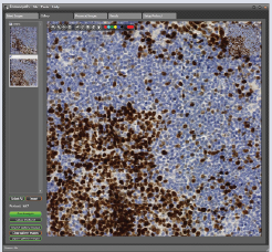 | 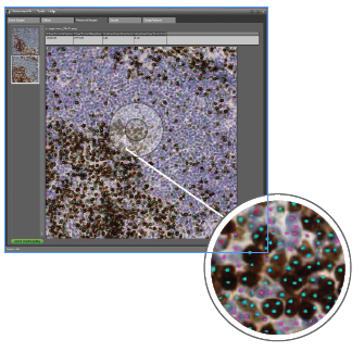 |
1. Load and select an image. Draw the area to be analyzed. | 2. Select one or more samples. Run analysis. | 3. Review and save results. |
The Immunopath system has been designed so that it is easy to setup simple protocols for the analysis of various samples and preperations, these are then applied using a simple 3 step process.
Image Analysis Examples
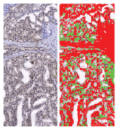 | 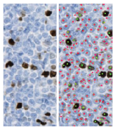 | 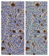 |
Sample stained with an antibody against PTEN. After running a field fraction analysis the area of positive cytoplasm stain is shown in green and the non-epithelial areas are red. | Based on the automated Immunopath analysis, the Ki-67-positive and negative nuclei are indicated by complementary coloured dots. | Formalin fixed paraffin embedded tumor tissue stained for Polo like kinase 1 (nuclei), counterstained with hematoxilin. |
Flexible protocol based image analysis for Whole Slide Images
Immunopath automises laborious and repetetive pathology tasks
More Information
Click here to download a brochure.
Use the contact form to request a demo version here.
Demonstration Video