PWS Ploidy Work Station
 | Automated Image based DNA Ploidy - Automatic high resolution scanning of monolayers
- Supports multiple slide scanning
- Automatic control of microscope, (navigation, focusing, image grabbing)
- Saves high quality images (TIF) and .nuc files
- Automatic segmentation and classification
|
DNA Ploidy is becoming an accepted prognostic marker. The DNA Ploidy test alone can be used to obtain prognostic information to determine treatment decisions in prostatic, urinary bladder, ovarian, endometrial and renal cell carcinoma among others. The test can also be an additive prognostic marker. DNA Ploidy provides a cost effective and objective way to assess the progression, make treatment decisions and to stratify patient groups. The system uses a research microscope, digital camera and a PC.
Prognostic markers
The main challenge for any oncologist is to predict the outcome of any chosen treatment for a cancer patient.
)
Epidemiologically and statistically, the probability of 5 year survival for treated cancer patients is estimated based on clinical staging, where survival is ranging from 60-95% in stage I to 0-15% in stage IV. With few exceptions, there is no method to predict if a single patient belongs to the non-surviving or surviving (good prognosis) group. A good prediction (a good prognostic marker) should allow the oncologist to choose the optimal treatment (or no treatment) for each patient, and hence either cure the patient with reduced long term complications and side effects, or improving life quality for those who can not be cured. Reducing the overall financial costs of treatment is also an added feature.
The main challenge for any oncologist is to predict the outcome of any chosen treatment for a cancer patient.
Epidemiologically and statistically, the probability of 5 year survival for treated cancer patients is estimated based on clinical staging, where survival is ranging from 60-95% in stage I to 0-15% in stage IV. With few exceptions, there is no method to predict if a single patient belongs to the non-surviving or surviving (good prognosis) group. A good prediction (a good prognostic marker) should allow the oncologist to choose the optimal treatment (or no treatment) for each patient, and hence either cure the patient with reduced long term complications and side effects, or improving life quality for those who can not be cured. Reducing the overall financial costs of treatment is also an added feature.
PWS - Ploidy Work Station system
The system comprises two elements, the PWS Grabber for microscope control and image capture and PWS Classifier for the analysis of the samples.
PWS Grabber
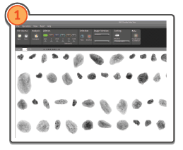
An application for automatic microscope scanning of monolayers. The PWS Grabber is employed to focus, grab, and segment cancer nuclei stained with the Feulgen-Schiff method. The absorbed stain is proportional to the DNA content in each nucleus.
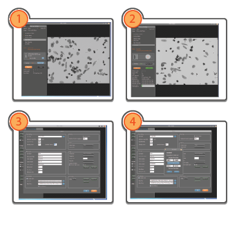 | Simple Operation 1.Slides and case ID are specified. 2. Adjust focus for slide 1. Set background position to an area without any objects and adjust light intensity. Adjust focus for slides in position 2-4 using the slider bar. 3.Controlled Setup Procedure. After logging in, tools and settings may be altered. 4. The options panel let’s you alter scan specifications, specify microscope system, camera and stage details, cell classification rules can be loaded, and positions to be scanned can be specified. Here are also possibilities to correct for the optical challenges like dark current, glare |
Main features of the software:
- Automatic high resolution scanning of monolayers
- Supports multiple slide scanning
- Automatic control of microscope, (navigation, focusing, image grabbing)
- Saves high quality images (TIF) and .nuc files
- Automatic segmentation and classification
Cell classification is completed using a rules set file, that is made based on results from manual editing. Different rules are used for different cell types and for monolayer and sections. The rules set makes it possible to store only the correctly segmented cells, and classify them in to galleries containing different cell types.
PWS Classifier
Automatic classification of cell types. PWS Classifier handles the images of the objects grabbed and segmented by the PWS Grabber.The software enables manual editing (sorting of objects), it displays DNA ploidy histograms, and performes calculations and statistics of selected objects. Finally, PWS Classifier enables manual and automatic DNA ploidy classification that could be used in diagnosis and prognosis of cancer patients.
 | 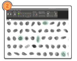 |
After opening a new case one may select the wanted image selection and start sorting the nuclei by selected parameter. | Nuclei are sorted into the galleries by area and the editing of each case by selecting nuclei may start. Misplaced nuclei can be put in other galleries. |
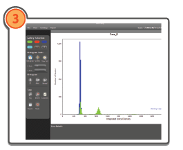 |  |
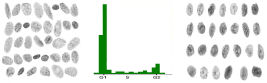
After editing the histogram can be opened. The nuclei in the different galleries are indicated by different colors. The selection and setting of the 2c peak is done. | Tags with statistical information of the selected peak may be added and it is possible to select and view the selected objects. The whole process can also be done automatically. |
 | 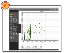 |
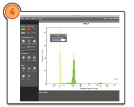
The results are added to a report where the histogram and the related statistical information is saved. | A scatter plot enables a plot with selected parameters. During setup it is possible to adjust for the optical features glare, dark current and diffraction. |
More information
Click here to download a brochure
Video showing system in operation click here.
The Room4 DNA ploidy system is intended for research use only.
System Hardware
The system is based on a research microscope and a high quality digital camera. It includes software and hardware to capture images from the monolayer and to produce a histogram of the DNA content of the cell nuclei enabling a DNA Ploidy classification to be made
Selected Publications on DNA Ploidy click here.