Products / Immunopath
Immunopath
Flexible, protocol-based image analysis for quantitative immunohistochemistry on whole slide images.
- Automates laborious, repetitive pathology tasks
- Full image analysis, with sampling options for data reduction
- Whole slide image support, plus import from standard imaging devices and common file formats
- Protocol-based analysis for repeatable, reliable results
- Runs on standard desktop PCs and laptops
Also available: Pathtool, a partner application with tools for grading microscopy images in pathology.
Why quantify IHC?
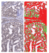
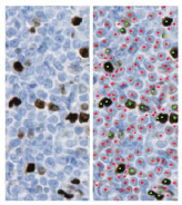
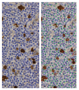
Reviewing immunohistochemistry slides by eye is slow and laborious. Counting cells or estimating the percentage of stain on a slide is not something most pathologists enjoy. Immunopath is a protocol-based tool that quickly completes cell counting and field fraction quantification on images from standard microscopy cameras and from whole slide imaging systems.
Immunohistochemistry (IHC) detects specific molecules on cells. Several diseases or disease subtypes can look alike under a microscope yet behave differently and need different treatment. One way to tell them apart is to detect marker molecules on the cells, using antibodies that find and attach to those markers. The antibodies can then be seen under the microscope and quantified. IHC is used widely in medicine, especially in cancer diagnosis and drug discovery.
The system
Immunopath provides quantitative information from IHC images captured with slide scanners and from single images captured with standard microscopy cameras.
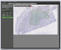
It is protocol-based. Protocols can be built from scratch or adapted from existing ones. Once a protocol is set up, the same image analysis steps run on every image and the results appear in a simple workflow. Protocols move easily between systems, so you can increase throughput and share analysis methods with colleagues.
A simple three-step process
- Load and select an image, then draw the area to analyse.
- Select one or more samples and run the analysis.
- Review and save the results.
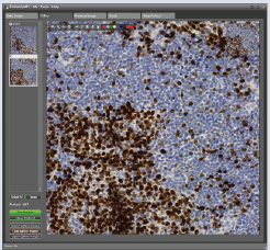
Image analysis examples